The Immune System 101
The immune system, unlike many other systems in the body, is a functional system comprised of molecules and immune cells that circulate throughout the body and occupy lymphoid tissues (Marieb & Hoehn, 2019, p. 782). This system is a combination of two intrinsic internal defense systems that work together to combat a slew of forces such as bacteria, fungi, diseases, infections, and cancer, providing us with a “resistance to disease” called immunity (p. 782). The lymphatic system—the structure of the immune system—consists of lymph, lymph nodes, and lymphatic vessels. Nutrients, gas, and waste are exchanged between the cells and tissues of the body through the blood and interstitial fluid, the fluid between tissues (p. 767).
Innate defenses: The first and second lines of defense
The body has two innate lines of defense; the mechanical barriers (i.e., the skin and mucosae) and cellular and chemical barriers work together to provide a rapid response to defend our bodies from harmful pathogens1 (Marieb & Hoehn, 2019, p. 782).
Surface barriers: Skin and mucosae. The skin and mucosae are our first line of defense within the innate defense system. The epidermis is highly keratinized, and it is this feature that makes our skin resistant to most weak acids, bases, bacterial enzymes, and toxins. Mucus membranes line all body cavities that are exposed to the exterior, that is, our digestive, respiratory, urinary, and reproductive tracts (Marieb & Hoehn, 2019, p. 782).
The skin and mucosae produce an assortment of protective chemicals such as acid, enzymes, mucin, and defensins. The acid mantle of our skin, the vaginal walls, and the stomach all function to inhibit bacterial growth. Enzymes function to destroy and digest bacteria. Mucin lines our respiratory tract and functions to trap debris and microorganisms and wash them into the stomach to be digested. Defensins are “broad spectrum antimicrobial peptides” found in mucous membranes and in our skin secretions (Marieb & Hoehn, 2019, p. 783). Defensins function to “control bacterial and fungal colonization” in areas where the surface area becomes breached (p. 783).
Internal defenses: Cells and chemicals. Cells and chemicals are our second line of defense within the innate defense system. The innate immune system uses pattern recognition receptors to detect potential microorganismal threats such as bacteria, viruses, parasites, and fungi (Marieb & Hoehn, 2019, p. 783). Of these receptors, the toll-like receptors (TLRs) are the most crucial in triggering immune responses in the body. There are eleven types of TLRs, each of which recognizes and attacks a different class of microorganism (p. 783).
Phagocyte cells within the connective tissue digest pathogens that penetrate the surface barriers of the immune system (Marieb & Hoehn, 2019, p. 784). The process of phagocytosis occurs in five stages. First, the phagocyte recognizes foreign microbes and debris via its receptors, then attaches itself to the pathogen through opsonins2 or antibodies. The phagocyte then forms a pseudopod that swallows the particle, enabling it to become a phagosome. This phagosome merges with a lysosome3 to form a phagolysosome. The lysosomal enzymes then digest the pathogens (p. 784).
Neutrophils, a type of phagocyte, are white blood cells that become phagocytic when they are met with infectious microbes. Free macrophages roam our tissues for “foreign invaders” while fixed macrophages inhabit specific organs and circulate through the tissues of the organ in search of debris (p. 784). Neutrophils and macrophages both destroy pathogens through phagocytosis; however, neutrophils also use defensins to help pierce the membrane of the pathogen. Neutrophils die rapidly in their defense; however, macrophages can survive multiple defense attacks against pathogens (p. 784).
Natural Killer (NK) cells are cells that “patrol” the body, circulating through blood and lymph. These cells, as one could derive from their name, are natural killers—they can attack cancer and virus cells before the adaptive immune system is called into action. NK cells do not discriminate against various kinds of cancers or viruses and are not phagocytic; rather, they directly aim at the target cell and induce the cell into apoptosis, or programmed cell death (Marieb & Hoehn, 2019, p. 785).
Inflammation and minor tissue injury. Inflammation is our immune system’s response to any tissue injury in the body (Marieb & Hoehn, 2019, p. 785). Inflammation functions to prevent the spread of any potentially hazardous agents to other surrounding tissues, assists in disposing of pathogens and other cellular debris, sets the stage for repair, and activates the adaptive immune system (p. 785).
Let’s say you get a minor cut on your hand—the inflammatory response initiates with mast cells4 triggering the release of inflammatory chemicals such as histamines5, kinins6, prostaglandins7, and cytokines8—a crucial foundation of maintaining tissue homeostasis in the body (Marieb & Hoehn, 2019, p. 785). These chemicals do three things: trigger vasodilation9, increase the permeability of the capillaries, and attract neutrophils, monocytes, and lymphocytes to the injury site (Larouche et al., 2018). Lymphatic tissues are home to lymphocytes and macrophages and provide the breeding ground for more lymphocytes (Marieb & Hoehn, 2019, p. 770).
Lymphatic capillaries create a tiny network between blood capillaries and tissue cells; they are highly permeable because their endothelial cells are loosely joined (Marieb & Hoehn, 2019, p. 768). Because their endothelial cells are loosely joined, collagen filaments help to hold the endothelial cells in place when the minivalves of the lymphatic capillaries open to receive interstitial fluid; these valves close when the pressure inside the capillary is greater than that outside (p. 768). The increased permeability of capillaries and venules local to the injury site allows exudate10 to flood into the surrounding tissues.
Clotting agents form a mesh that helps to prevent the spread of bacteria and provide a scaffolding for tissue repair (Marieb & Hoehn, 2019, p. 786). Here, one might experience one or multiple symptoms of acute inflammation such as pain, heat, swelling, and redness at the injury site, as well as impairment of function (Marieb & Hoehn, 2019, p. 787). The increased temperature at the site of injury increases the metabolic rate of the cells to promote healing (Larouche et al., 2018). See Figure 1 for a complete chart of the events of inflammation.
Simultaneously, once the inflammation process has begun, phagocytes mobilize into the affected area. This process begins with leukocytosis, the increase of white blood cells in the body in response to an injury. Leukocytosis-inducing factors11 are released into the blood, hence triggering neutrophils to flow into the blood from red bone marrow within only a couple of hours of the injury (Marieb & Hoehn, 2019, p. 786). Next, phagocytes begin gravitating to the inner walls of capillaries and venules—a process called margination. The endothelial cells surrounding become inflamed and bud cell adhesion molecules (CAMs), which signal to the body a metaphorical target of the injury site. Neutrophils begin to bind with these cell adhesion molecules, triggering more CAMs to bud (p. 786). Diapedesis occurs as a result; neutrophils begin to flatten and squeeze out of the capillaries at the injury site. Inflammatory chemicals continue to flood the area, homing neutrophils further into the damaged area (See Figure 2). Macrophages from the lymphatic tissues act as a garbage disposal of inflammatory debris, pathogens, and dead tissue (p. 770, 786). These macrophages are essential in the healing process because not only do they clear out the “debris” of the injury, they secrete more cytokines and growth factors (such as IL-1), and this in and of itself promotes the proliferation of fibroblasts and new epithelial cells (Larouche et al., 2018). Lymphatic vessels draw excess protein-filled interstitial fluid—now called lymph—back into the bloodstream for circulation (Marieb & Hoehn, 2019, p. 767). When a tissue becomes inflamed, the lymphatic capillaries grow to absorb larger particles such as pathogens, cancer cells, and dead cell debris. Like veins, lymphatic capillaries lead to larger channels—lymphatic vessels, lymphatic trunks, and lymphatic ducts. Because the lymphatic system lacks a pump, like the heart for the circulatory system, the lymphatic system relies on changes in thoracic pressure and skeletal muscle movements to circulate lymph into the lymph nodes (p. 769). Lymph nodes scatter themselves along the lymph vessels in the body, with macrophages cleansing the lymph before returning it to the blood (p. 771). In addition to lymph nodes, the white pulp of the spleen is where the majority of the immune functions occur—cleaning the body of pathogens by “recycling the breakdown products of red blood cells” and storing monocytes for later use; the “pulp color” is reflective of the actual appearance of the spleen and has no other significance (p. 774).
Depending on the type of tissue injured and the severity of the injury, tissue repair occurs in one of two ways: regeneration or fibrosis. In regeneration, destroyed tissue is replaced with the same kind of tissue; in fibrosis, destroyed tissue is replaced with scar tissue, a dense connective tissue12 (Marieb & Hoehn, 2019, p. 143). In a nonextensive wound of the skin, like a papercut, cytokines within the inflammatory response set the stage for clotting and thus, for fibroblasts13 to proliferate and produce growth factors and collagen (Larouche et al., 2018). When healing of the injury is complete, the cells undergo apoptosis, leaving macrophages and phagocytes to remove the dead cells. Epithelial cells then begin to multiply over the granulation tissue, a fragile tissue rich with capillaries (Marieb & Hoehn, 2019, pp. 143-144). The wounded area is then pulled together slowly through the fibrotic tissue, regenerating under the scab (clot) that formed over the wound. The scab will fall off once the wound is healed and the epithelium has regenerated. However, monocytes from the initial immune response could linger at the injury site for up to a few weeks (Marieb & Hoehn, 2019, p. 144; Larouche et al., 2018).
Antimicrobial proteins and contraction of influenza. Antimicrobial proteins are proteins that work in accordance with our innate defenses by attacking microbes and/or hindering their reproduction. Since viruses do not have the cellular capability to generate adenosine triphosphate or synthesize proteins, they invade the body’s tissues, taking over the cells so they can reproduce (Marieb & Hoehn, 2019, p. 788). Interferons (IFNs) are small proteins secreted by infected cells that work to protect uninfected cells. First, the virus enters the cell and begins to reproduce, triggering the IFN genes into action. The infected cell begins to produce IFNs, which bind and trigger the cell’s genes for antiviral proteins. The antiviral proteins then work in accordance to hinder viral reproduction (p. 788). See Figure 3 for a visual representation of this mechanism.
Mucosa-associated lymphoid tissues (MALT) are located within the mucous membranes of the body, functioning to help protect us against pathogens. The largest of these is the tonsils, which remove any pathogens from inhaled air (pp. 774-775). However, sometimes a pathogen or cancer cell passes through the lymph node and can circulate freely throughout the body (Marieb & Hoehn, 2019, p. 769). Seasonal influenza is one common example of a virus that can overtake the body. Influenza is first categorized by A, B, C, or D; for this example, we will explore influenza A virus (IAV), as it is most widely spread across humans and animals. The virus enters the body via an infected particle from another person through an opening such as our nose, eyes, or mouth. The molecular structure of IAV—that is, the structure of its hemagglutinin (HA) and neuraminidase (NA) proteins—is related to its severity and “immune evasion”; the way HA attaches itself is a major key in B cell antibody production in the body and thus becomes the determinant in viral mutations. This high variability is what permits IAVs to evade our “immune surveillance” and, as a result, leads to seasonal influenza (Chen et al., 2018). IAV enters the cell and begins to reproduce, triggering the IFNs genes into action, triggering the infected cell to produce IFNs, further triggering the cell’s genes for antiviral proteins (p. 788).
Once the virus is detected by the body, the innate immune system kicks in, triggering inflammatory responses in the body. The flood of cytokines and histamines throughout the body might lead one to experience increased mucus production, difficulty breathing as the respiratory system contracts, and pain (Marieb & Hoehn, 2019, p. 785). Leukocytes and macrophages, when exposed to pathogens, release pyrogens, a chemical that acts on the neurons in the hypothalamus to raise the body’s temperature, leading one to experience a fever (Marieb & Hoehn, 2019, p. 789). A fever also activates the liver and spleen to remove iron and zinc, which “makes them less available to support bacterial growth” (p. 789). While one might feel terrible symptoms, the recruitment of the antibody14 army is already underway.
The complement system (complement) is a system of about twenty plasma proteins that work together to decimate foreign bodies. When active, complement stimulates inflammatory chemicals throughout the body and kills bacteria. Essentially, complement enhances the innate and adaptive defense systems within the body (Marieb & Hoehn, 2019, p. 788). There are three ways complement is activated. In the classic pathway, antibodies bind to the pathogen, activating their ability to bind to complement. In the lectin pathway, lectins15 bind to sugars on the surface of the invading microorganism, activating their ability to bind to complement. Lastly, in the alternative pathway, C3, one of the complement proteins, interacts with the microorganism on its surface (p. 788). All of the of these pathways intersect at C3, splitting it into C3a and C3b; this splitting is what initiates and “enhances inflammation, promotes phagocytosis, and causes cell lysis16” (Marieb & Hoehn, 2019, p. 788).
Adaptive defenses: The third line of defense
The adaptive immune system is a fairly precise mechanism for attacking and eliminating pathogens (Marieb & Hoehn, 2019, p. 790). The adaptive system gets triggered into action through exposure to foreign substances, or antigens, and, unlike the innate immune system, takes a much longer time to come into effect fully (p. 790). Additionally, the adaptive immune system differs from the innate immune system in that it is specific, targeting identified pathogens, is not restricted to the infection or injury site, and after initial exposure, can recognize previously encountered pathogens, viruses, and bacteria and attack them (p. 791).
There are two mechanisms within the adaptive immune system: humoral immunity and cellular immunity. Humoral immunity, also known as antibody-mediated immunity, consists of the antibodies present in the humors, fluids such as blood and lymph, of the body; these antibodies freely circulate throughout the body and bind to extracellular targets such as viruses, bacteria, and other toxins (p. 790). Cellular immunity, or cell-mediated immunity, is active when lymphocytes17 defend the body by attacking a variety of cells infected with pathogens, bacteria, viruses, cancer, and foreign grafts (p. 790).
Antigens. Antigens are large, foreign, complex molecules that become the target of the adaptive immune system and act to mobilize it. There are two types of antigens: complete or incomplete. Complete antigens have immunogenicity; that is, they can trigger lymphocytes to reproduce. There are only specific parts of an antigen that are immunogenic; these antigenic determinants dictate whether or not the body recognizes the substance as foreign. For example, plastic, a large, simple molecule, is often used in artificial implants because it has “identical, regularly repeating units” and therefore has “little or no immunogenicity” (Marieb & Hoehn, 2019, p. 791). Because of this, plastic is not seen as foreign and will not be rejected by the body. Complete antigens can react to the antibodies released within the body and activated lymphocytes (p. 791). Pollen, bacteria, fungi, and viruses are all immunogenic and examples of complete antigens.
Incomplete antigens, also known as haptens, are small molecules that bind with the body’s proteins. When this happens, the body’s adaptive immune system can recognize this pairing as foreign and attack the body rather than protect it. Haptens have reactivity but lack immunogenicity (Marieb & Hoehn, 2019, p. 791). Chemicals such as poison ivy, animal dander, detergents, cosmetics, and household products are some examples of incomplete antigens (p. 791).
Lymphocytes and antigen-presenting cells. Within the adaptive immune system are three key cells: B lymphocytes (B cells), T lymphocytes (T cells), and antigen-presenting cells (APCs). B cells are those of which moderate humoral immunity. T cells are lymphocytes that do not produce antibodies and make up the cellular “arm” of the adaptive immune system (Marieb & Hoehn, 2019, p. 792). APCs have auxiliary responsibilities, such as helping T cells recognize their antigens; they do not respond to antigens the way B or T cells do (p. 792).
Both B and T cells originate in red bone marrow. B cells mature within the bone marrow while T cells mature within the thymus, a primary lymphoid organ(18) which lacks B cells and thus does not directly mount attacks against antigens (Marieb & Hoehn, 2019, p. 776). As T cells mature, they become immunocompetent and “display a unique type of receptor on their surface” (p. 792). This receptor is what allows them to identify and bind to antigens. They also learn self-tolerance; that is, they become unresponsive to their self-antigens(19) and therefore do not attack their own cells (p. 792). During maturation, lymphocytes are educated through positive and negative selection. In positive selection, “only T cells with receptors that can recognize self-MHC proteins(20) survive”; T cells that do not pass this test undergo apoptosis (Marieb & Hoehn, 2019, p. 794). Negative selection allows only T cells that “do not recognize self-antigens displayed on self-MHC” to survive—all others undergo apoptosis (p. 794). This maturation process takes about two to three days. Then, naïve B and T cells, cells that have not been exposed to an antigen, are released from the primary lymphoid organs to seed secondary lymphoid organs21. This circulation of B and T cells increases their probability of coming into contact with antigens throughout the body, lymphocytes, and APCs. B and T cells are most likely to encounter antigens in the lymph nodes or spleen, where they will bind with the specific antigen, a process called clonal selection. The lymphocyte activates and multiplies, forming an “army” of like cells called clones. A majority of clones become effector cells, working to fight the foreign invaders; however, some of these cells become memory cells, which can identify and rapidly respond to subsequent encounters with familiar antigens (pp. 792-793). It is important to note that our genes are responsible for which pathogens, viruses, and other foreign substances our body can detect and fight, not our antigens; antigens are only responsible for determining which lymphocytes will be called on to mount the attack. See Figure 4 for a complete diagram of the lymphocyte development, maturation, and activation process.
APCs, as mentioned, are responsible for “delivering” antigens to their “compatible” cells—dendritic cells22, macrophages, and B lymphocytes.
Humoral immune response. A humoral immune response occurs when a B cell encounters an antigen (Marieb & Hoehn, 2019, p. 796). B cells become activated when the antigen binds to the receptors on its surface; the cell then undergoes clonal selection and is proliferated and differentiated into effector cells and memory cells. Here, a majority of B cells become effector cells, plasma cells, which secrete antibodies (p. 796). Plasma cells can secrete antibodies at about 2000 molecules per second. However, they are only active for about four or five days before they die. The remaining cells become memory cells with the ability to mount a humoral attack nearly immediately when encountering a familiar antigen (p. 796).
The primary immune response is the initial response to an antigen encounter, which occurs about three to six days after the initial exposure. Once mobilization has begun, plasma antibodies rise, peaking around ten days after initial exposure. The secondary immune response occurs when the body re-encounters a familiar antigen; the body calls on its immunological memory to mount an attack against the antigen. This response takes about two to three days to take effect within the body (Marieb & Hoehn, 2019, p. 797).
Active immunity is when B cells encounter an antigen and produce antibodies to defend against it. This happens either naturally, when, for example, one gets an infection, or artificially, when one is vaccinated. Many vaccines contain weakened, dead, or components of pathogens. Weakened antigens within a vaccine can “provide functional antigenic determinants that are both immunogenic and reactive” (Marieb & Hoehn, 2019, pp. 797-798) and additionally, “spare” one of the symptoms that generally accompany the disease or infection that would come as a side effect of the primary immune response (p. 798). Passive humoral immunity occurs when ready-made antibodies are presented to the body. Here, B cells are not being tested by antigens and therefore, immunological memory does not occur, and as such, protection ceases when the antibodies degrade in the body (p. 798).
Immunoglobulins (Igs), or antibodies, form the gamma globulin aspect of blood proteins (Marieb & Hoehn, 2019, p. 798). While antibodies differ based on their antigen counterpart, all antibodies have four polypeptide chains to form a molecule called the antibody monomer. Two of these chains, the heavy (H) chains, are identical to one another; the other two light (L) chains are also identical to one another. They are generally half as long as the H chains. Within each chain is a variable (V) region and constant (C) region at opposing ends. C regions are nearly the same across all antibodies, while V regions vary by antigen interaction. The V regions of each of the four chains come together to form two antigen-binding sites. The C regions, or stem of the antibody monomer, are also known as the effector regions of the monomer, which specify the chemicals or cells that the antibody can bind to and how it will function to detect and destroy antigens. There are five major classes of immunoglobulins: IgM, IgA, IgD, IgG, IgE (p. 799). In the example of IAV, the most important antibody in recovering from the infection is the IgG monomer because it is abundant in the body and is present in both the primary and secondary immune responses (Chen et al., 2018). See Table 1 for the characteristics of each immunoglobulin class.
Antibodies function in many ways. While they cannot directly destroy antigens, they can “flag” antigens as targets to be destroyed, an interaction called an antigen-antibody complex. There are three ways an antigen-antibody complex can become activated, leading to phagocytosis; in neutralization, antibodies “block specific sites” on viruses and toxins so that the antigen cannot bind to tissue cell receptors (Marieb & Hoehn, 2019, 800). Antibodies also cause agglutination, or particle clumping, when the antigen-antibody complexes cross-link, forming large, lattice-like particles. Agglutinated particles that are soluble are more easily phagocytized—a process called precipitation. The antigen-antibody complex can also trigger complement activation, which not only enhances phagocytosis but encourages inflammation and cell lysis (p. 800).
Cellular immune response. A cellular immune response occurs when a T cell encounters an antigen (Marieb & Hoehn, 2019, p. 801). T cells can both kill other cells that are infected, cancerous, abnormal, or foreign and release chemicals to trigger immune responses. There are two main groups of T cells: CD4 and CD8. CD4 and CD8 are both glycoproteins that, when activated, develop into one of three groups of effector cells: helper T (TH) cells, regulatory T (TREG) cells, and cytotoxic T (TC) cells (p. 801).
A majority of CD4 cells develop into TH cells. These cells activate other T cells, B cells, and macrophages, and lead the adaptive immune responses (Marieb & Hoehn, 2019, p. 802). They are essential in the adaptive immune response. TH cells release cytokines, which trigger rapid cell division and enhance the innate immune system by mobilizing macrophages and lymphocytes (p. 806). TH1 cells trigger inflammation, the activation of macrophages, and differentiation of TC cells. TH2 cells are responsible for activating immune responses against parasitic worms and promoting allergies. TH17 cells “merge” the innate and adaptive immune responses through the release of IL-17, an interleukin that triggers inflammatory responses against many autoimmune diseases (p. 806).
When activated, some CD4 cells become TREG cells, which function to moderate the adaptive immune responses (p. 802). These cells moderate the immune system by releasing cytokines23 which inhibit the immune response (p. 807). Activated CD8 cells develop into TC cells, which attack and destroy foreign cells in the body (p. 802). TC cells target viruses, bacteria, parasites, cancerous cells, and foreign (transplanted) cells. Like NK cells, TC cells circulate throughout the body surveilling and examining cells for recognizable markers; the difference, however, is that NK cells “check to make sure each cell has ‘identity flags’ . . . whereas TC cells check ‘identity flags’ to see if they look the way they are supposed to” (Marieb & Hoehn, 2019, p. 807). Some activated CD4 and CD8 cells can also develop into memory T cells that can help regulate immune responses when re-exposed to a familiar antigen (p. 802).
Unlike B cells, T cells are not able to recognize free or natural antigens—they can only identify and react to “processed fragments of protein antigens displayed on surfaces of body cells” (Marieb & Hoehn, 2019, p. 803). There are two classes of MHC proteins that T cells can recognize: class I and class II. Class I MHC proteins are present on the surface of almost all cells of the body save red blood cells. Class I MHC proteins have a facet that can bind with a protein fragment of an endogenous antigen, that is, an antigen of which the fragments of proteins are synthesized within the cell (p. 803). When protein-digesting enzymes begin to digest cytoplasmic proteins, protein fragments enter the endoplasmic reticulum (ER) of the cell, where newly synthesized class I MHC proteins bind to peptides between eight and nine amino acids long. These class I MHC proteins are then transported to the cell surface. Class I MHC proteins essential function to activate CD8 cells and relay information about infectious, abnormal, or cancerous cells within the body to TC cells (p. 803). Separately, class II MHC proteins are generally only found on cell surfaces presenting antigens to CD4 cells (e.g., dendritic cells, macrophages, B cells). Similar to class I MHC proteins, class II MHC proteins are also synthesized within the ER of the cell. The peptides that class II MHC proteins bind to can be about fourteen to seventeen amino acids long and are derived from exogenous antigens, or antigens that have come from outside of the cell and been absorbed into the cell (p. 803). Exogenous antigens that have been engulfed by a cell are broken down by phagolysosomes, then bound to class II MHC proteins before being transported to the cell surface; this then signals a “call for help” to other CD4 cells (p. 803).
Both CD4 and CD8 cells, similar to B cells, undergo an “educational process” during maturity; this process is called MHC restriction. In MHC restriction, CD4 cells “learn” their restriction of “binding antigens only on class II MHC proteins . . . typically only displayed on APC surfaces” (Marieb & Hoehn, 2019, p. 804). CD8 cells, which become activated by antigens on the surface of class I MHC proteins, can also be found on the surfaces of APCs and, when active, begin scouting for the same antigen on other cells throughout the body (p. 804). When dendritic cells detect IAV, for example, the conventional dendritic cells move from the lungs to the lymph nodes, where they present the antigen to T lymphocytes (Chen et al., 2018). Dendritic cells have this ability because they can “engulf” virus-infected or cancerous cells, enabling them to display antigens to both class I and class II MHC proteins (Marieb & Hoehn, 2019, p. 805). The dendritic cells then dismantle the viral protein into immune peptides, which are then transported to the cell’s ER to bind with class I MHC. After they join, the merged class I MHC is transported to the cell surface so that TC can recognize them (Chen et al., 2018).
T cells are only able to be activated by APCs (Marieb & Hoehn, 2019, p. 805). First, T cell antigen receptors (TCRs) bind with an antigen-MHC complex on an APC’s surface. The TCR will both recognize the MHC protein and the antigen that it displays. Before being able to proliferate, the T cell must be co-stimulated by a co-stimulatory signal, a molecule on the surface of the APC of damaged or infected tissues (p. 805). If the T cell were to bind with an antigen without this co-stimulatory signal, the cell would become anergic, or unresponsive and tolerant to the antigen, and thus, this co-stimulatory signal is crucial for activated T cell effectiveness in the immune response (p. 805). TC assists in the production of cytokines and effector molecules, which are key players in inhibiting the virus from multiplying. Chen et al. state, though, that “viral proteins degraded in endosomes/lysosomes are associated with MHC class II molecule,” and because of this, they are recognized by TH cells, triggering B cell proliferation and maturation, a process that takes about a week after antigen exposure (Chen et al., 2018; Marien & Hoehn, 2019, 805)
Using the example of IAV again, once antibody production begins, antibodies will circulate freely throughout the body, binding to IAV (Marieb & Hoehn, 2019, p. 790). After antibodies bind with IAV, the molecule now becomes active in its ability to bind with complement. The complement C3 then splits into C3a and C3b, promoting phagocytosis and cell lysis, leading to an eventual recovery in the otherwise “healthy” person between seven and thirty days after initial exposure (p. 780).
References
Akdis, M., Verhagen, J., Taylor, A., Karamloo, F., Karagiannidis, C., Crameri, R., Thunberg, S., Deniz, G., Valenta, R., Fiebig, H., Kegel, C., Disch, R., Schmidt-Weber, C. B., Blaser, K., & Akdis, C. A. (2004). Immune responses in healthy and allergic individuals are characterized by a fine balance between allergen-specific T regulatory 1 and T helper 2 cells. The Journal of experimental medicine, 199(11), 1567–1575. https://doi.org/10.1084/jem.20032058
Chen, X., Liu, S., Goraya, M. U., Maarouf, M., Huang, S., & Chen, J. L. (2018). Host Immune Response to Influenza A Virus Infection. Frontiers in immunology, 9, 320. https://doi.org/10.3389/fimmu.2018.00320
Larouche, J., Sheoran, S., Maruyama, K., & Martino, M. M. (2018). Immune Regulation of Skin Wound Healing: Mechanisms and Novel Therapeutic Targets. Advances in wound care, 7(7), 209–231. https://doi.org/10.1089/wound.2017.0761
Marieb, E.N., and Hoehn, K. (2019). Human anatomy and physiology (11th ed.). Hoboken: Pearson Education, Inc.
Yu, W., Freeland, D., & Nadeau, K. C. (2016). Food allergy: immune mechanisms, diagnosis and immunotherapy. Nature reviews. Immunology, 16(12), 751–765. https://doi.org/10.1038/nri.2016.111
Definitions
Pathogens are harmful or disease-causing microorganisms (Marieb & Hoehn, 2019, p. 782).
Opsonins are molecules that coat the foreign invading microorganism, providing a means for macrophages and neutrophils to attach themselves and destroy the cell (Marieb & Hoehn, 2019, p. 789).
Lysosomes are organelles that live in cell membranes and contain activated digestive enzymes; they are abundant in phagocytes (Marieb & Hoehn, 2019, p. 86).
Mast cells detect foreign microorganisms and orchestrate the inflammatory responses against them; their cytoplasm has secretory granules of inflammatory-mediating chemicals, such as heparin (an anticoagulant), histamine (a chemical that makes capillaries leaky), and proteases (protein-degrading enzymes) (Marieb & Hoehn, 2019, p. 129).
Histamines are an inflammatory chemical within the body that function to increase vasodilation of arterioles, influencing hyperemia to occur and thus, allowing exudate to flood the injury site (Marieb & Hoehn, 2019, p. 785).
Kinins, like histamines, trigger an inflammatory response of vasodilation and hyperemia; however, kinins induce leukocytes and neutrophils to release lysosomal enzymes (Marieb & Hoehn, 2019, p. 785). Kinins also induce pain (p. 785).
Prostaglandins, like kinins, trigger an inflammatory response of vasodilation and hyperemia, induce pain, and occasionally can act as an anti-inflammatory (Marieb & Hoehn, 2019, p. 785).
Cytokines, also known as complements, are bloodborne proteins that lyse microbes, increase phagocytosis, and function to intensify inflammatory responses (Marieb & Hoehn, 2019, p. 785).
Vasodilation occurs when the lumen of blood vessels increases as the smooth muscle relaxes (Marieb & Hoehn, 2019, p. 709).
Exudate is a bodily fluid that contains clotting agents and antibodies (Marieb & Hoehn, 2019, p. 785).
Leukocytosis-inducing factors are chemicals that are released by injured cells (Marieb & Hoehn, 2019, p. 786).
Dense connective tissue is fibrous and withstands great tension, stress, and stretching (Marieb & Hoehn, 2019, pp. 132-133).
Fibroblasts are immature connective tissue cells that are actively mitotic, that is, they rapidly divide and replicate (Marieb & Hoehn, 2019, p. 129).
Antibodies are water-soluble proteins produced by the adaptive immune system to fight off pathogens (Marieb & Hoehn, 2019, p. 788).
Lectins are water-soluble proteins produced by the innate immune system to recognize pathogens (Marieb & Hoehn, 2019, p. 788).
Cell lysis is the process by which a cell disintegrates. It begins when C3b binds to the surface of the target cell. This binding stimulates the insertion of complement proteins known as MAC, or membrane attack complex. MAC stabilizes this hole in the membrane, which allows water to enter the cell, causing lysis (Marieb & Hoehn, 2019, p. 789).
Lymphocytes are a type of leukocyte, or white blood cell; few are found in the bloodstream; rather, they are mostly found in the lymphoid tissues such as the lymph nodes and spleen (Marieb & Hoehn, 2019, p. 653).
Bone marrow and the thymus (Marieb & Hoehn, 2019, p. 792).
Self-antigens are a variety of protein molecules that speckle the surface of all of the cells in our bodies; they are neither foreign nor antigenic to each person; however, they are antigenic to the nonself (Marieb & Hoehn, 2019, p. 792).
MHC (major histocompatibility complex) proteins are glycoproteins that live on the cell surface and help the cell identify as self or nonself (Marieb & Hoehn, 2019, p. 791).
All other lymphoid organs (Marieb & Hoehn, 2019, p. 792).
Dendritic cells are mobile fighting cells with long extensions perfect for capturing antigens. They internalize antigens through phagocytosis and deliver them to T cells (Marieb & Hoehn, 2019, p. 794).
Cytokines are groups of molecules that influence cell development, differentiation, and immune responses. Cytokines include interferons and interleukin 1 (IL-1), a glycoprotein which promotes T cell activation, inflammation and fever, and interleukin 2 (IL-2), a glycoprotein which triggers both B and T cell proliferation, TREG development, and activates NK cells (Marieb & Hoehn, 2019, p. 805).
Tables
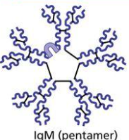
IgM
This is the first class of immunoglobulins. During the primary immune response, plasma cells secrete IgM, which activates complement. IgM can exist in both monomer and pentamer forms. The monomer version “receives” antigens on the surface of B cells. The pentamer version circulates throughout blood plasma. Because there are many antigen-binding sites, IgM is a strong agglutinating-particle-clumping-agent.
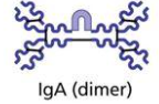
IgA
IgA is a dimer found in secretions such as sweat and saliva. Also known as Secretory IgA, this dimer helps to prevent pathogens from attaching to the epithelial cell surface. IgA is limited and found in plasma.
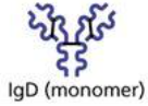
IgD
The IgD monomer is found on the surface of cells where it acts as a receptor for antigens.
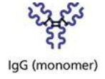
IgG
The monomer IgG is the most abundant immunoglobulin in the body and is the primary antibody in both primary and secondary immune responses. IgG activates complement to protect against foreign substances, viruses, bacteria, and other toxins as it circulates throughout the blood and lymph. IgG is a monomer that is involved in passive immunity, where, for example, it crosses from the placenta of the mother to her fetus.